
INTRODUCTION: Overview of Surgical Technique
“There is no excuse today for the surgeon to [first] learn on the patient.”
-- Dr. William J. Mayo, 1927
The three-dimensional microanatomy of the human temporal bone is complex and challenging. Mastery of cadaveric temporal bone dissection prior to live otologic surgery is a central tenet of training in otolaryngology and otology/neurotology. In no other speciality can one practice the most fundamental of microsurgical procedures in a model that so closely resembles the live patient. Fortunately, the most critical structures within and adjacent to the temporal bone are quite resilient following cadaveric specimen preparation; dissection of the dura, sigmoid sinus, facial nerve, ossicles, labyrinth and carotid artery in the temporal bone lab closely simulates operating room dissection.
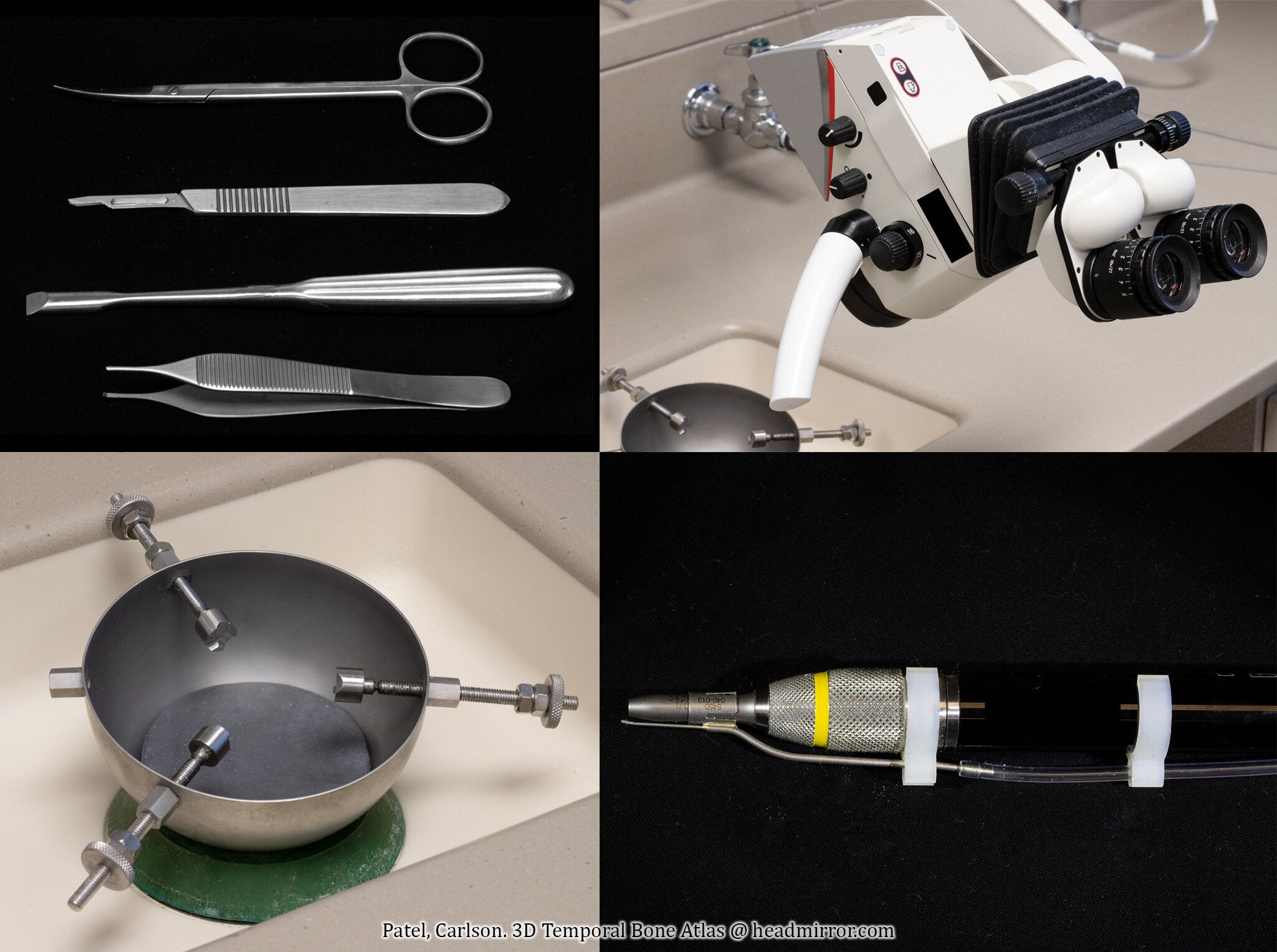

Practice like you play. Basic instrumentation including an otologic drill, an operating microscope, and a limited set of otologic microinstruments are all that is required to rehearse the full breadth of otologic and neurotologic microsurgery in the temporal bone lab. As well, dissection can be approached systematically with a “lateral to medial” approach around or through structures of interest that allows a host of procedures to be performed on a single specimen. For example, a sequence comprising intact canal wall mastoidectomy, followed by facial recess with cochleostomy, endolymphatic sac decompression, labyrinthectomy, identification and decompression of the internal auditory canal (translabyrinthine exposure), posterior canal wall removal, cochleoectomy (for transotic exposure), and full decompression with subsequent posterior mobilization of the facial nerve (for transcochlear exposure) allows for at least 7 distinct procedures to be performed on one temporal bone specimen.
However, it is not always feasible for trainees to be regularly paired with experienced mentors for hours in the dissection lab. Nor can invaluable specimens be wasted simply because the trainee did not have an adequate visual guide or dissection manual. This atlas, paired with available texts, videos, podcasts, and descriptions of surgical technique, aims to provide a helpful guide for unsupervised temporal bone dissection at all levels of surgical competency. We hope you enjoy!
Neil S. Patel, MD and Matthew L. Carlson, MD
Reference:
Mayo WJ. Medical education for the general practitioner. JAMA 1927;88(18):1377-1379